Smooth Muscle Diagram / The Role Of Vascular Smooth Muscle Cells In The Physiology And Pathophysiology Of Blood Vessels Intechopen - In this video i have shown the simplest way of drawing muscle drawing.
Smooth Muscle Diagram / The Role Of Vascular Smooth Muscle Cells In The Physiology And Pathophysiology Of Blood Vessels Intechopen - In this video i have shown the simplest way of drawing muscle drawing.. 1024x840 draw a labelled diagram of a smooth muscle diagram of smooth. Hand | definition, anatomy, bones, diagram, & facts. As in cardiac muscle cells, the configuration of the nuclear membranes in smooth muscle cells changes during contraction and. Cardiac, skeletal and smooth muscles are the three types of muscles found in the human body. Smooth muscle images stock photos vectors shutterstock.
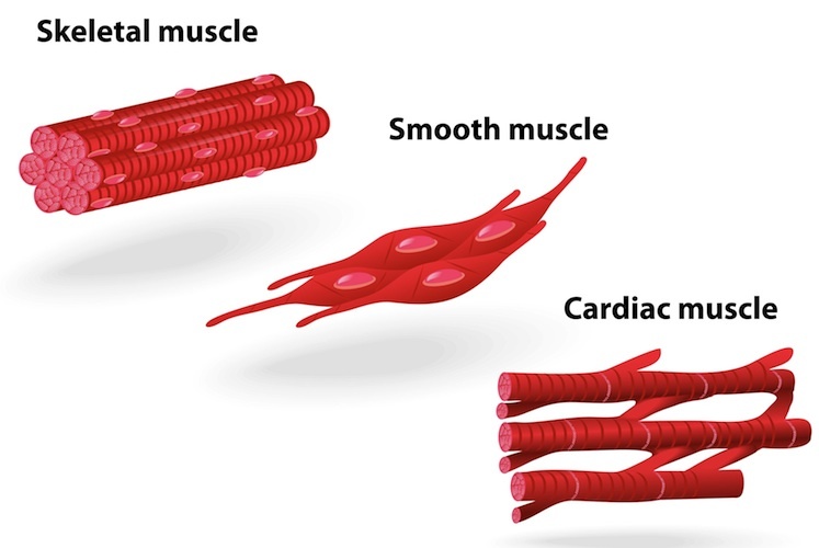
Smooth muscle has a fusiform shape, which resembles a football or spindle. It is the pen diagram of skeletal, smooth and cardiac muscle for class 10, 11 and 12. *smooth muscle* the cardiovascular, gastrointestinal, genitourinary, and respiratory systems are smooth muscle thus subserves all internal, involuntary functions, except the movements of breathing. Smooth muscle histology and diagram (inlet). 1024x840 draw a labelled diagram of a smooth muscle diagram of smooth.
Smooth Muscle Cells Cardiovascular Research Oxford Academic from static.primary.prod.gcms.the-infra.com Hand | definition, anatomy, bones, diagram, & facts. Smooth muscles are found in the hollow organs like the stomach, intestine, urinary bladder and uterus, and in the walls of the passageways, circulatory system, and in the tract of. Vascular smooth muscle refers to the particular type of smooth muscle found within, and composing the majority of the wall of blood vessels. • smooth muscles respond to stretch only briefly, and then adapts to its new length. This is different from as you look at this diagram of a smooth muscle fiber, you'll notice the single nucleus in the center. They work automatically without you being aware of them. Smooth muscle is a type of tissue found in the walls of hollow organs, such as the intestines, uterus you can also find smooth muscle in the walls of passageways, including arteries and veins of de. Smooth muscle tissue is also known as visceral muscle tissue.
This diagram depicts muscle of the body diagrams 744×1054 with parts and labels.
They work automatically without you being aware of them. Smooth muscles, cardiac muscles and skeletal muscles. It is layered in a distinctive pattern of circular layers. This diagram depicts muscle of the body diagrams 744×1054 with parts and labels. 12 photos of the smooth muscle diagram. Diagram of artery with smooth muscle identification. Hand | definition, anatomy, bones, diagram, & facts. Smooth muscle (factors affecting activation, general properties, source of cytosolic ca2+, structure, muscle cells). Smooth muscle and cardiac muscle move to facilitate body functions like heartbeats and digestion. Cardiac, skeletal and smooth muscles are the three types of muscles found in the human body. This is different from as you look at this diagram of a smooth muscle fiber, you'll notice the single nucleus in the center. There are three types of muscles in the body: *smooth muscle* the cardiovascular, gastrointestinal, genitourinary, and respiratory systems are smooth muscle thus subserves all internal, involuntary functions, except the movements of breathing.
In this video i have shown the simplest way of drawing muscle drawing. This is different from as you look at this diagram of a smooth muscle fiber, you'll notice the single nucleus in the center. Vascular smooth muscle contracts, blood vessels become narrower and less blood flows through them. By ning zhou, shaunrick stoll. Smooth muscle (factors affecting activation, general properties, source of cytosolic ca2+, structure, muscle cells).
The Three Types Of Muscle Tissue Of Human Body Diagram Stock Illustration Illustration Of Blood Actin 193729908 from thumbs.dreamstime.com *smooth muscle* the cardiovascular, gastrointestinal, genitourinary, and respiratory systems are smooth muscle thus subserves all internal, involuntary functions, except the movements of breathing. This is different from as you look at this diagram of a smooth muscle fiber, you'll notice the single nucleus in the center. Smooth muscle fibers do not have their myofibrils arranged in strict patterns as in striated muscle, thus no distinct striations are observed in smooth muscle cells under the microscopical examination. Vascular smooth muscle contracts, blood vessels become narrower and less blood flows through them. Diagram of smooth muscle contraction, smooth cardiac and skeletal muscle diagram, smooth muscle cell diagram, smooth muscle cell picture. By ning zhou, shaunrick stoll. They work automatically without you being aware of them. 1024x840 draw a labelled diagram of a smooth muscle diagram of smooth.
Smooth muscle and cardiac muscle move to facilitate body functions like heartbeats and digestion. Smooth muscle images stock photos vectors shutterstock. Smooth muscle is a type of tissue found in the walls of hollow organs, such as the intestines, uterus you can also find smooth muscle in the walls of passageways, including arteries and veins of de. It is layered in a distinctive pattern of circular layers. By ning zhou, shaunrick stoll. Vascular smooth muscle is the type of smooth muscle that makes up most of the walls of blood vessels. It is the pen diagram of skeletal, smooth and cardiac muscle for class 10, 11 and 12. Smooth muscle, muscle that shows no cross stripes under microscopic magnification. It is divided into two subgroups; This is different from as you look at this diagram of a smooth muscle fiber, you'll notice the single nucleus in the center. They work automatically without you being aware of them. In this video i have shown the simplest way of drawing muscle drawing. Smooth muscles, cardiac muscles and skeletal muscles.
Smooth muscle images stock photos vectors shutterstock. Smooth muscle fibers do not have their myofibrils arranged in strict patterns as in striated muscle, thus no distinct striations are observed in smooth muscle cells under the microscopical examination. Vascular smooth muscle contracts, blood vessels become narrower and less blood flows through them. It is divided into two subgroups; Smooth muscle tissue is also known as visceral muscle tissue.
Muscle Structure Muscle Under The Microscope Science Learning Hub from static.sciencelearn.org.nz Vascular smooth muscle refers to the particular type of smooth muscle found within, and composing the majority of the wall of blood vessels. Here presented 43+ smooth muscle drawing images for free to download, print or share. Smooth muscles, cardiac muscles and skeletal muscles. Smooth muscle has a fusiform shape, which resembles a football or spindle. 12 photos of the smooth muscle diagram. There are three types of muscles in the body: Vascular smooth muscle is the type of smooth muscle that makes up most of the walls of blood vessels. As in cardiac muscle cells, the configuration of the nuclear membranes in smooth muscle cells changes during contraction and.
Smooth muscle, muscle that shows no cross stripes under microscopic magnification.
Smooth muscle and cardiac muscle move to facilitate body functions like heartbeats and digestion. • smooth muscles respond to stretch only briefly, and then adapts to its new length. Learn vocabulary, terms and more with flashcards, games and other study tools. Smooth muscle histology and diagram (inlet). Smooth muscle has a fusiform shape, which resembles a football or spindle. Here presented 43+ smooth muscle drawing images for free to download, print or share. This diagram depicts muscle of the body diagrams 744×1054 with parts and labels. Smooth muscles, cardiac muscles and skeletal muscles. Hand | definition, anatomy, bones, diagram, & facts. Vascular smooth muscle contracts, blood vessels become narrower and less blood flows through them. In this video i have shown the simplest way of drawing muscle drawing. As in cardiac muscle cells, the configuration of the nuclear membranes in smooth muscle cells changes during contraction and. Maximus ilium, sacrum, coccyx and lumbodorsal fascia iliotibial tract and femur extension and lateral rotation at the hip.


Posting Komentar
0 Komentar