The 19 Muscles Of The Foot - What Preventative Exercises Can Strengthen Intrinsic Foot ... - The ultrasound appearances of ankle and foot ligaments, tendons, and nerves are similar to those in other parts of the body.
The 19 Muscles Of The Foot - What Preventative Exercises Can Strengthen Intrinsic Foot ... - The ultrasound appearances of ankle and foot ligaments, tendons, and nerves are similar to those in other parts of the body.. The first part is and introduction to the muscles of the foot and covers the dorsal group of muscles and some aspects of innervation. The ultrasound appearances of ankle and foot ligaments, tendons, and nerves are similar to those in other parts of the body. The dorsal aponeurosis of the toes supports the effect of the dorsal foot muscles by redirecting the force line of their tendons to. Flexion of 4 lesser toes at metatarsophalangeal, proximal & distal interphalangeal joints inversion of foot plantar flexion of ankle. Flexor hallucis longus tendon transfer to the dorsum of the foot and release of the flexor digitorum longus and brevis tendons at the base of each toe.
Foot muscle forces & deformities. 26.20 superficial intrinsic muscles of the sole right foot, plantar view. They are considered voluntary muscles. The other 19 muscles are referred to as intrinsic muscles of the foot and act only within the foot. First layer • the first layer of muscles is the most superficial to the sole, and is located immediately underneath the plantar fascia.
Tennis Ball Self Massage - Your Definitive Trigger Point ... from ignorelimits.com Learn vocabulary, terms and more with flashcards, games and other study tools. Using an elastic band, the athlete loops the band around one foot and steps on the other end; Start studying muscles of the foot. Extensor digitorum longus (extension of last 4 toes and. This means that the little toe can only be extended by the extensor digitorum longus muscle only. Insertions of the extrinsic foot muscle tendons on the plantar surface of the foot. The short and long muscles of the foot serve as synergists. The foot of these creeping animals is extremely muscular, penetrated by nerves, and capable of generating one, two, or four laterally adjacent contraction waves.
10.19 (a) pattern of peripheral sensory innervation in the right lower limb.
Learn vocabulary, terms and more with flashcards, games and other study tools. There are 29 muscles associated with the human foot. A thick, flat muscle located beneath the gastrocnemius, and together the form the calf of the leg; A generous moment arm of these muscles about the midfoot. The first part is and introduction to the muscles of the foot and covers the dorsal group of muscles and some aspects of innervation. Goes to medial cuneiform bone/ medial aspect of the base of 1st metatarsal. There are 2 neurovascular planes between the muscle layers of the sole What is foot muscles?/foot muscles acting on the foot can be divided into two distinct groups; • fourth layer ( fig. Flexion of 4 lesser toes at metatarsophalangeal, proximal & distal interphalangeal joints inversion of foot plantar flexion of ankle. They are considered voluntary muscles. Explore the muscles of the foot in this complete guide! There are many ligaments in the foot.
Several of the pims span the la in parallel with the plantar. A thick, flat muscle located beneath the gastrocnemius, and together the form the calf of the leg; It acts with the gastrocnemius to. 26.3 joints of the foot right foot with talocrural joint in plantar flexion. Contrary to expectations, the intrinsic foot muscles contribute minimally to supporting the arch of the foot during walking and running.
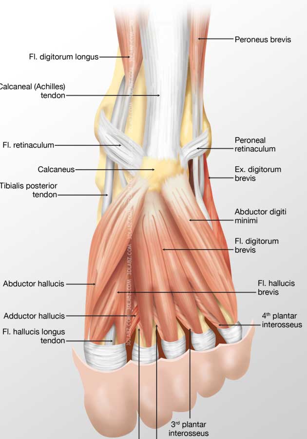
Foot Dorsal Muscles 3D Illustration | Price from www.3dlabz.com Start studying muscles of the foot. The short and long muscles of the foot serve as synergists. Foot muscle forces & deformities. The muscles covered in this article serve various. The muscles acting on the foot can be divided into two distinct groups; The muscles mainly responsible for movement of the foot are the anterior and posterior pedal retractors. (from schuenke m, schulte e fig. Neurovascular planes of the sole:
Word doc of all foot muscles, attachments and movements foot muscles anterior muscles tibialis anterior (dorsiflexion and inversion) comes from lateral condyle.
The first part is and introduction to the muscles of the foot and covers the dorsal group of muscles and some aspects of innervation. Insertions of the extrinsic foot muscle tendons on the plantar surface of the foot. First layer • the first layer of muscles is the most superficial to the sole, and is located immediately underneath the plantar fascia. The muscles acting on the foot • the muscles acting on the foot can be divided into two distinct groups; Several of the pims span the la in parallel with the plantar. Flexion of 4 lesser toes at metatarsophalangeal, proximal & distal interphalangeal joints inversion of foot plantar flexion of ankle. The dorsal muscles of the foot. 26.19 intrinsic muscles of the dorsum right foot, dorsal view. The muscles acting on the foot can be divided into two distinct groups; Arises from the tibia and fibula, it extends to the heel; The muscles covered in this article serve various. Muscle layers of the sole of the foot. The ultrasound appearances of ankle and foot ligaments, tendons, and nerves are similar to those in other parts of the body.
Using an elastic band, the athlete loops the band around one foot and steps on the other end; It acts with the gastrocnemius to. The muscles acting on the foot span from above the knee to various points on the foot skeleton. Those of the medial plantar region are connected with the great toe, and corrrespond with those of the thumb; • fourth layer ( fig.
Several things about Anatomy of the Hip Muscles | Foot ... from i.pinimg.com (10 foot/ankle and 19 intrinsic) ten of these muscles originate outside of the foot itself but the other 19 muscles are referred to as intrinsic muscles of the foot and act only within the foot. The muscles mainly responsible for movement of the foot are the anterior and posterior pedal retractors. The interosseous muscles of the foot are muscles found near the metatarsal bones that help to control the toes. Neurovascular planes of the sole: There are 2 neurovascular planes between the muscle layers of the sole They are considered voluntary muscles. Contrary to expectations, the intrinsic foot muscles contribute minimally to supporting the arch of the foot during walking and running. Learn vocabulary, terms and more with flashcards, games and other study tools.
The skeleton of the foot is often subdivided, based on functional and clinical 10.16 the short muscles of the right foot from the plantar view.
• fourth layer ( fig. However, these muscles do influence our ability to produce forward propulsion from one stride into the next, highlighting their role in bipedal locomotion. First layer • the first layer of muscles is the most superficial to the sole, and is located immediately underneath the plantar fascia. The dorsal muscles of the foot. Muscles of the ankle and foot. The interosseous muscles of the foot are muscles found near the metatarsal bones that help to control the toes. Flexion of 4 lesser toes at metatarsophalangeal, proximal & distal interphalangeal joints inversion of foot plantar flexion of ankle. (a) the insertions of the flexor digitorum longus, flexor hallucis longus and little attention has been paid to the clinical assessment of intrinsic foot muscles in the musculoskeletal injury literature apart from few specific. 10.19 (a) pattern of peripheral sensory innervation in the right lower limb. There are 29 muscles associated with the human foot. The muscles mainly responsible for movement of the foot are the anterior and posterior pedal retractors. The muscles of the anterior compartment of the lower leg are generally responsible for dorsiflexion, and the muscles of the posterior compartment of the lower leg are generally responsible the lateral and medial muscles in both compartments invert, evert, and rotate the foot. A thick, flat muscle located beneath the gastrocnemius, and together the form the calf of the leg;


Posting Komentar
0 Komentar